Diagnostic Imaging: Unveiling Insights with Precision
Diagnostic imaging is a fundamental component of modern medicine, enabling healthcare professionals to visualize the internal structures of the body and diagnose a wide range of medical conditions accurately. The field of diagnostic imaging utilizes advanced technologies and imaging modalities to provide detailed and high-quality images for interpretation by radiologists and other specialists. Let’s explore the key aspects of diagnostic imaging:
- X-rays: X-ray imaging is one of the oldest and most widely used diagnostic techniques. It involves exposing the body to a small amount of ionizing radiation to capture images of bones, organs, and tissues. X-rays help identify fractures, lung diseases, infections, and certain abnormalities.
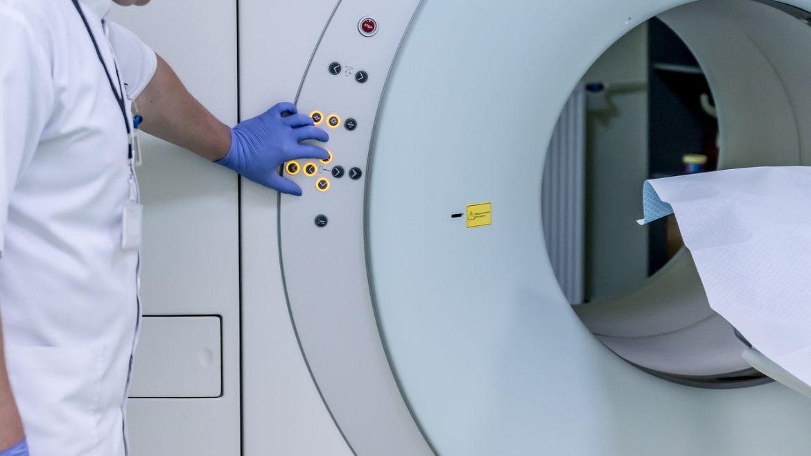
- Computed Tomography (CT): CT scans utilize a combination of X-rays and computer technology to create detailed cross-sectional images of the body. CT scans provide more detailed information than traditional X-rays, allowing for the visualization of soft tissues, blood vessels, and internal organs. They are especially useful for diagnosing complex conditions, such as tumours, cardiovascular diseases, and neurological disorders.
- Magnetic Resonance Imaging (MRI): MRI uses a strong magnetic field and radio waves to generate detailed images of the body’s organs and tissues. It is particularly effective in visualizing soft tissues, muscles, ligaments, and the central nervous system. MRI scans are valuable in diagnosing conditions such as brain and spinal cord injuries, joint abnormalities, and tumours.
- Ultrasound Imaging: Ultrasound imaging uses high-frequency sound waves to create real-time images of the body’s internal structures. It is commonly used for prenatal care, as it allows visualization of the developing fetus. Ultrasound is also utilized to assess the abdominal organs, blood vessels, and musculoskeletal system.
- Nuclear Medicine: Nuclear medicine involves the administration of small amounts of radioactive materials to the patient, which are then detected by specialized cameras to create images of specific organs and evaluate their functions. It is particularly useful in diagnosing and treating conditions related to the thyroid, bones, heart, and certain types of cancers.
- Positron Emission Tomography (PET): PET imaging combines nuclear medicine and computerized tomography to provide detailed metabolic information about the body. It is primarily used in cancer diagnosis and staging, as well as in evaluating brain disorders, cardiac conditions, and certain neurological conditions.
The Integration and Impact:
The integration of cardiology services and diagnostic imaging is pivotal in delivering comprehensive and precise healthcare. Cardiologists rely on accurate imaging techniques to assess heart health and plan appropriate interventions, while radiologists collaborate with cardiologists to interpret cardiovascular images effectively. The synergy between these two fields significantly improves patient care and outcomes.
Technological Innovations:
Cardiology services and diagnostic imaging continually evolve with technological advancements. Innovative technologies such as 3D imaging, artificial intelligence (AI), and telemedicine have revolutionized these fields. 3D imaging provides an enhanced visualization of cardiac structures and assists in surgical planning. AI algorithms aid in the early detection of cardiovascular diseases and streamline image interpretation. Telemedicine facilitates remote consultations, enabling patients in remote areas to access specialized cardiology services and diagnostic imaging.